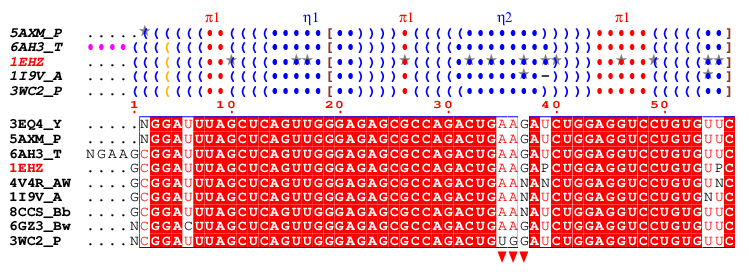
- The secondary structure elements in Dot-Bracket Notation (DBN, see the ViennaRNA documentation
 ) are derived from PDB files using X3DNA-DSSR (Liu et al., 2015) and are presented above the sequence block. They are coloured and labelled as follows: ) are derived from PDB files using X3DNA-DSSR (Liu et al., 2015) and are presented above the sequence block. They are coloured and labelled as follows:
- blue
() indicate regular base pairs
- orange
() are wobble pairs
- maroon
<>, {}, [] are pseudo-knots
- blue dots, labeled
η, indicate hairpin loops
- cyan dots, labeled
β, indicate bulges
- green dots, labeled
ι, indicate internal loops
- red dots, labeled
π, indicate junctions
- purple dots, labelled
α, indicate non-loop single stranded segments
- Hyphens (
–) indicate gaps and grey stars (★) highlight modified bases, if present.
- Below the DBN block is the multiple sequence alignment, the default colouring of which is based on a percentage calculation of strict identity for each residue column. Users can set a threshold (named Global Score - 70% by default) that distinguishes between low and high similarity:
- If the similarity score of a residue column exceeds this threshold, the residues are displayed in red with a blue frame.
- Columns with scores below the threshold are displayed in black.
- Regardless of the threshold value, residues with strict identity are always displayed as white on a red background.
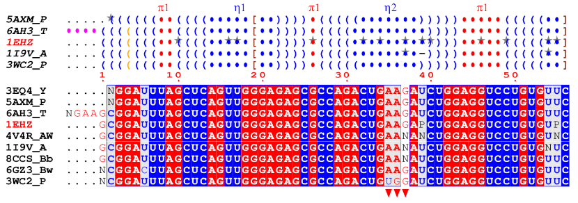
- Another colouring scheme is also possible, based on chemical properties (Colour by residues chemical properties option in the Special commands and characters section), which produces the following figure:
- Here, purine residues (
A and G) are written in white on a red background if they are strictly conserved. They are written in red on a grey background if there is >70% similarity (by default), and on a white background otherwise. The same scheme applies to pyrimidine residues (C, T, U), with red being substituted with blue. The remaining characters are in black.
- Finally, users can add custom markers as desired. In this example, three red triangles have been placed below the sequence block to indicate the position of the anticodon within the tRNAPhe sequences.
• Lu, X.J., Bussemaker, H.J., and Olson, W.K. (2015) DSSR: an integrated software tool for dissecting the spatial structure of RNA. Nucleic Acids Res., 43(21):e142
|