NESPript is in its final stage of development. Consequently, there may be a few minor bugs remaining. If you experience any problems, please email us at espript@ibcp.fr with a detailed description of the issue. Rest assured that we will do our utmost to fix it as quickly as possible.
What is NESPript?
NESPript, 'Nucleic acids Easy Sequencing in PostScript', is a Web server designed for the representation of multiple alignments of nucleic acid sequences, including the visualization of their secondary structures in Dot-Bracket Notation (DBN) when available.
A comparable service is available for proteins through our ESPript server.
Key features:
NESPript produces a PostScript, PDF, PNG or TIFF file containing aligned sequences with graphical enhancements.
The main input is a file of pre-aligned sequences in Clustal, FASTA or MultAlin format.
The program calculates a similarity score for each residue and colors the alignment accordingly.
Secondary structure elements can be added to the multiple sequence alignment using PDB/CIF/DSSR files or automated search.
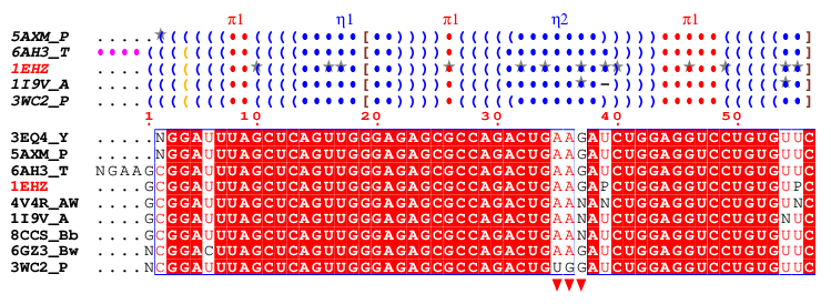
A typical figure produced by NESPript is shown below. It was generated from the alignment file clustal-1EHZ.aln which contains tRNAPhe sequences. For more details, please refer to Example #2 on our Tutorials & Examples page.
The secondary structure elements in DBN (see the ViennaRNA documentation ) are derived from PDB files using the program X3DNA-DSSR and are presented above the sequence block. They are coloured and labelled as follows:
blue () indicate regular base pairs
orange () are wobble pairs
maroon<>, {}, [] are pseudo-knots
blue dots, labeled η, indicate hairpin loops
cyan dots, labeled β, indicate bulges
green dots, labeled ι, indicate internal loops
red dots, labeled π, indicate junctions
purple dots, labelled α, indicate non-loop single stranded segments
Hyphens (–) indicate gaps and grey stars (★) highlight modified bases, if present.
Below the DBN block is the multiple sequence alignment, the default colouring of which is based on a percentage calculation of strict identity for each residue column. Users can set a threshold that distinguishes between low and high similarity:
If the similarity score of a residue column exceeds this threshold, the residues are displayed in red with a blue frame.
Columns with scores below the threshold are displayed in black.
Regardless of the threshold value, residues with strict identity are always displayed as white on a red background.
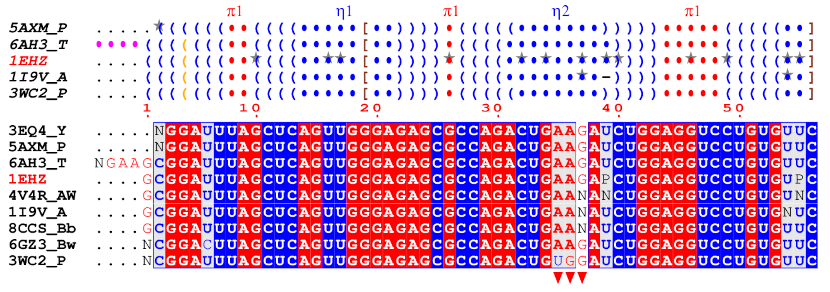
Another colouring scheme is also possible, based on chemical properties, which produces the following figure:
Here, purine residues (A and G) are written in white on a red background if they are strictly conserved. They are written in red on a grey background if there is >70% similarity (by default), and on a white background otherwise. The same scheme applies to pyrimidine residues (C, T, U), with red being substituted with blue. The remaining characters are in black.
Finally, users can add custom markers as desired. In this example, three red triangles have been placed below the sequence block to indicate the position of the anticodon within the tRNAPhe sequences.
Presentation of NESPript at the RNA Collaborative Seminar Series
NESPript is provided as a service to the scientific community and can be used free of charge for research and educational purposes without restriction.
We would appreciate it if you would use the appropriate citation when publishing data produced with NESPript.
Do not hesitate to contact us (espript@ibcp.fr) if you need any further information or if you need some help with NESPript.
The authors make no warranties regarding the correctness of the data. Their responsibility is limited to applying best efforts in providing the most reliable and accurate service. The authors are not responsible for the use of the results, data or information obtained from this server. This Web server is hosted on the CNRS network and governed by French law. By accessing this website or using all or part of its functions, users formally accept the application of French law. This website does not use tracking cookies or cookies that collect personal data for marketing or analytical purposes (see our 'Cookie Policy'). This website uses an encrypted connection (HTTPS). The IP address of the client computer and the date and time of the connection are processed for usage statistics. No other data is collected and no data is transferred to third party partners or sites.